A mushroom looks simple from a distance — a cap on a stem, pushed up out of the ground overnight. Look closer and that simplicity dissolves. Every visible mushroom is the temporary reproductive organ of a much larger organism, and each of its parts has a precise name, a developmental history, and a role in the fungus’s strategy for survival and reproduction.
Learning the anatomy of a mushroom is not an academic exercise. The features described here are exactly the ones mycologists use to tell species apart — including the difference between an edible mushroom and a deadly one. Understanding the structure is the foundation of everything else: identification, classification, ecology, and the biology of how these organisms actually live.
This guide walks through the full structure of a typical gilled mushroom, part by part, and explains what each feature tells you.
What You Are Actually Looking At
The first thing to understand is that the mushroom is not the organism. It is the fruiting body — the structure the fungus produces to make and release spores, in the same way an apple is the fruit of a tree rather than the tree itself.
The bulk of the fungus lives out of sight, as a network of microscopic threads called mycelium, threaded through soil, wood, or other substrate. For most of its life the fungus exists only as this network. When conditions are right — usually a combination of temperature, moisture, and nutritional status — the mycelium channels resources into building a fruiting body, pushing it up into the air where its spores can be caught by wind, water, or animals.
Everything visible above ground, then, is in service of one goal: producing and dispersing spores. The architecture of the mushroom is the architecture of a spore-delivery system.
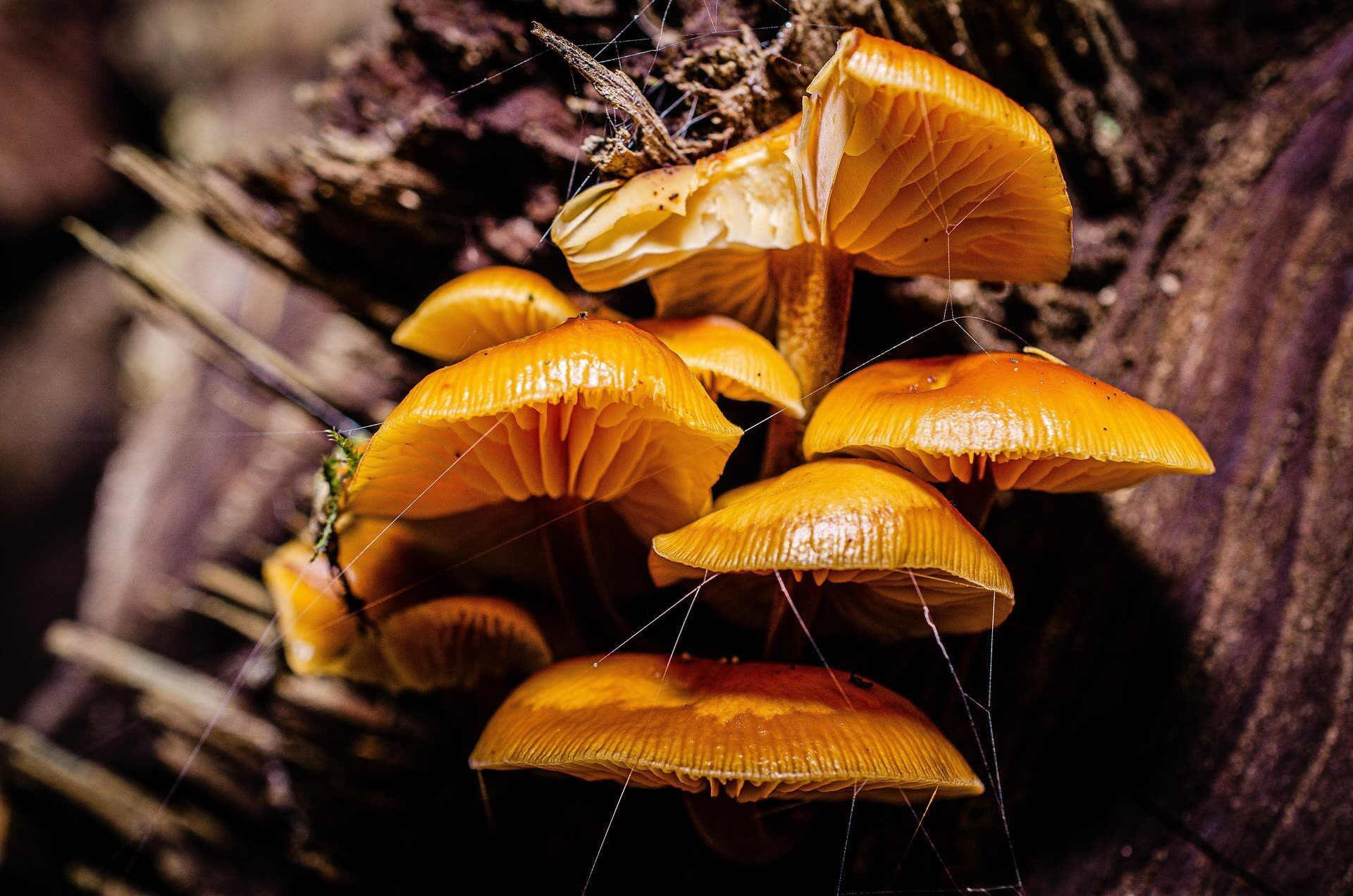
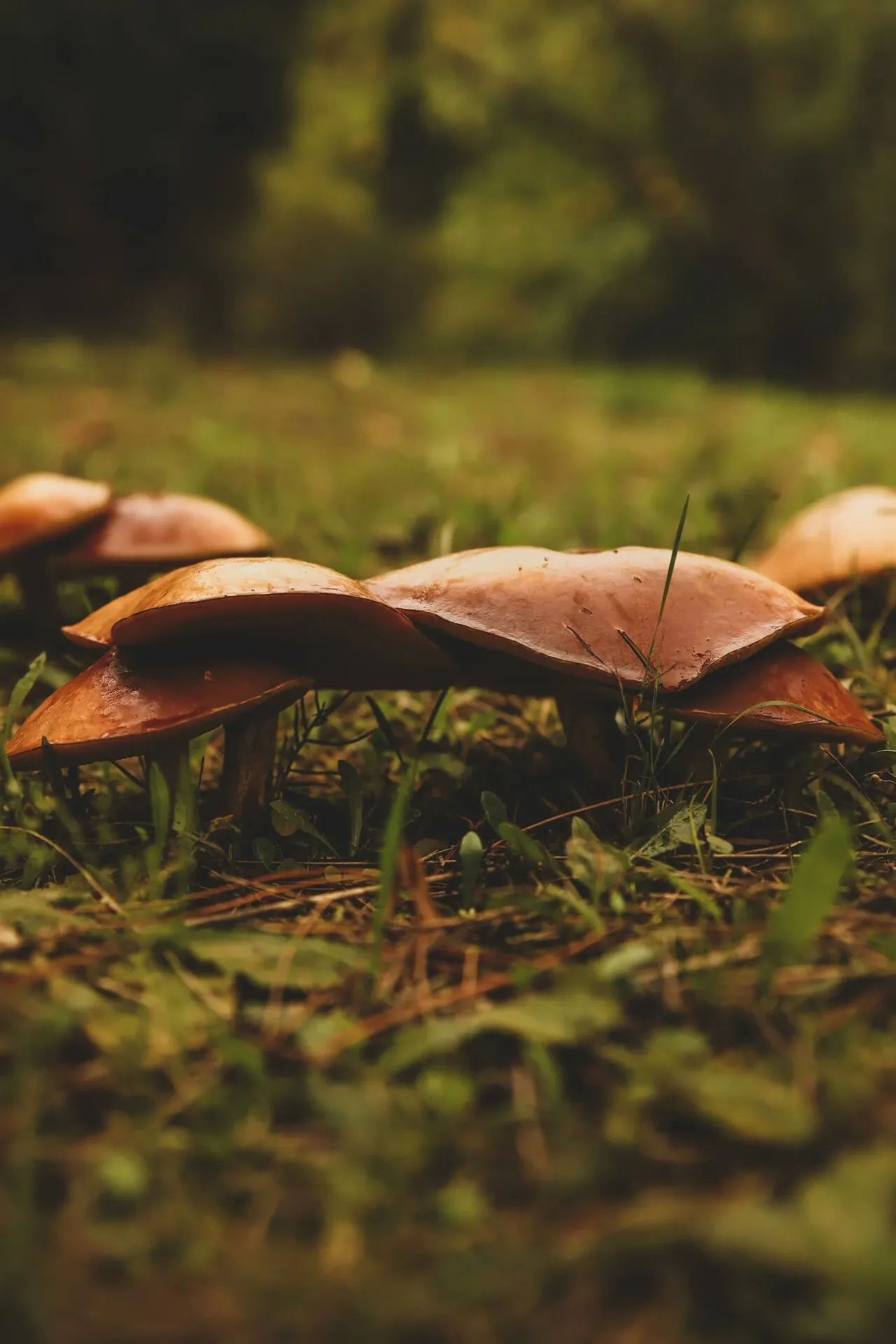
The Cap (Pileus)
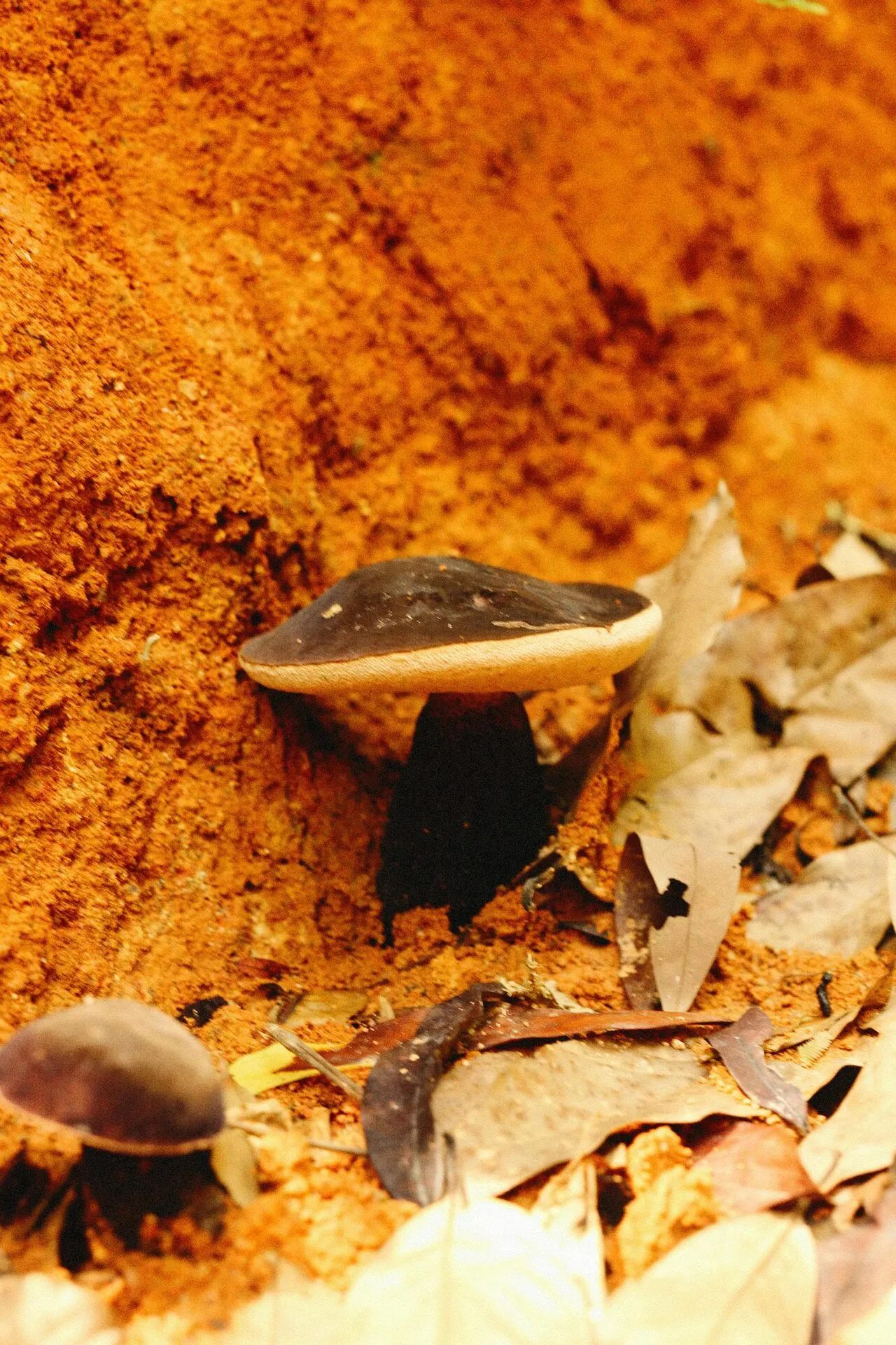
The cap, technically the pileus, is the most conspicuous part of the mushroom. Its primary job is to protect the spore-producing surface underneath and to position that surface at the right height and angle for spore release.
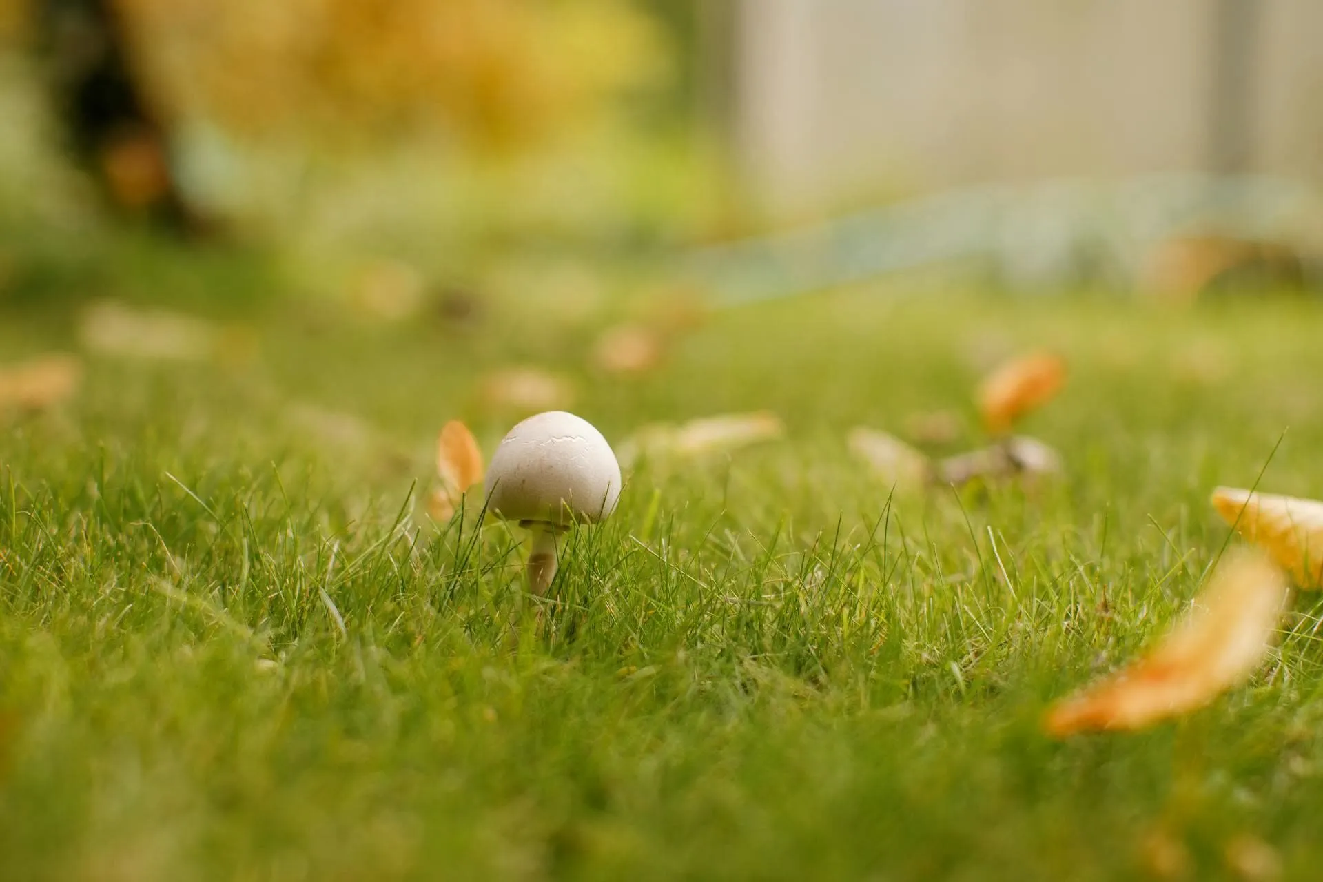
Caps vary enormously in shape, and the shape changes as the mushroom matures. Many begin convex or even spherical, protecting the developing gills, and flatten or become funnel-shaped with age. The progression from a rounded “button” to a flattened or upturned mature cap is itself an identification clue, because the rate and form of this change differ between species.
The surface of the cap carries information too. It may be smooth, scaly, fibrous, sticky, or covered in patches — remnants of tissue from earlier in development. Color matters, but with an important caveat: cap color in many species changes with age, moisture, and light exposure, so it is among the least reliable single features for identification.
A feature that deserves particular attention in psilocybin mushrooms is bruising. Many Psilocybe species bruise blue when handled or damaged, the result of psilocin oxidizing on contact with air. This bluing is suggestive but not definitive — it is one feature among many, and some non-psychoactive species also discolor.
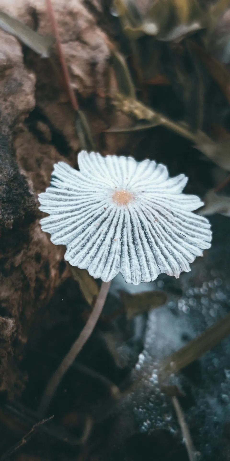
The Gills (Lamellae)
Turn a typical mushroom over and you find the gills — properly, the lamellae — radiating out from the stem to the cap edge. This is the spore factory. The gills are lined with a fertile layer called the hymenium, densely packed with the microscopic cells that produce and eject spores.
The architecture here is about surface area. By folding the fertile tissue into dozens or hundreds of thin vertical plates, the mushroom packs an enormous spore-producing surface into a compact cap. A single common mushroom can release billions of spores over its short life.
How the gills attach to the stem is one of the most useful identification features in all of mycology. Gills may run down the stem (decurrent), attach squarely to it (adnate), notch before reaching it (adnexed), or stop short and not touch it at all (free). These attachment types are consistent within species and visible to the naked eye, which is why field guides emphasize them.
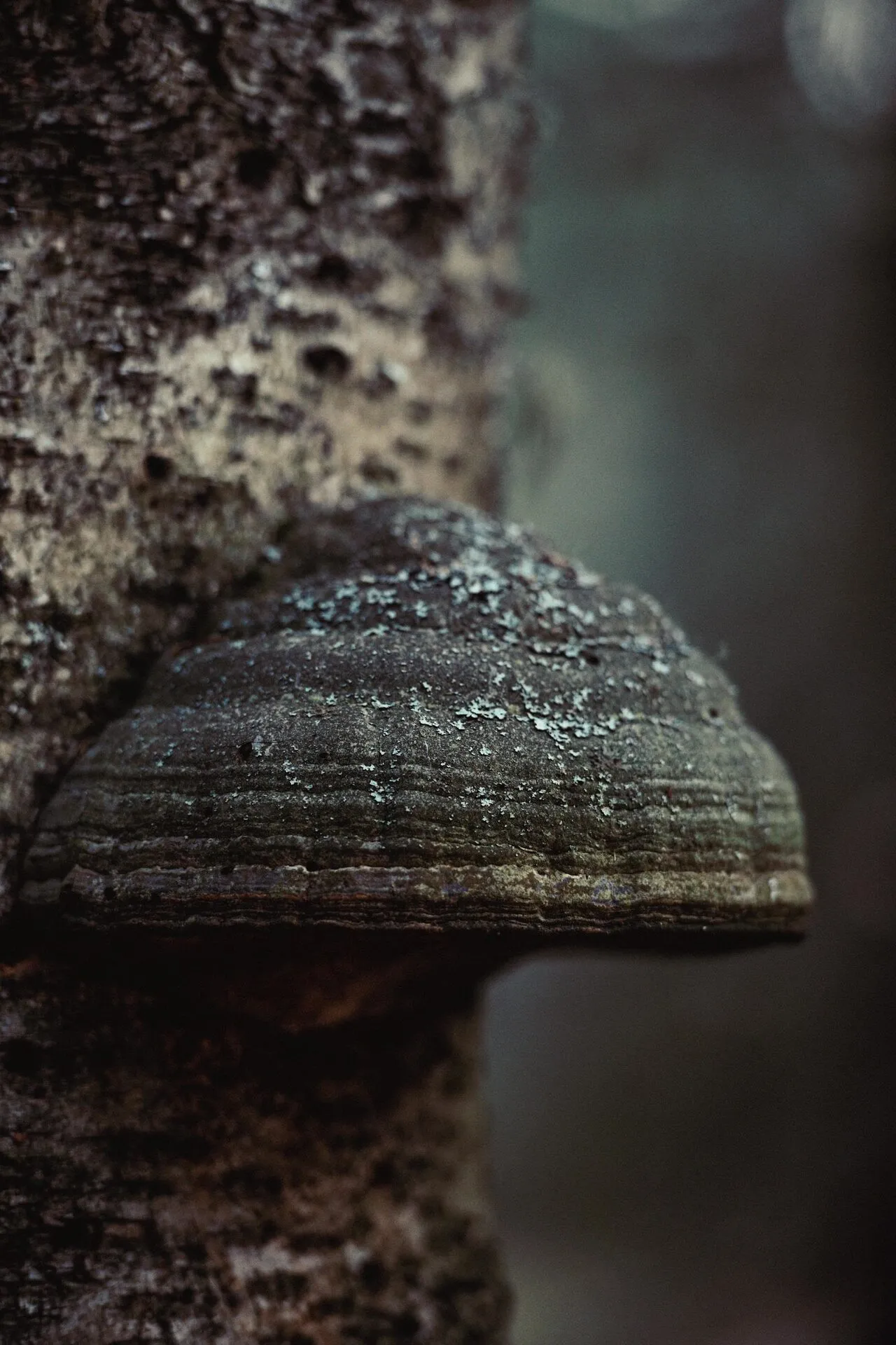
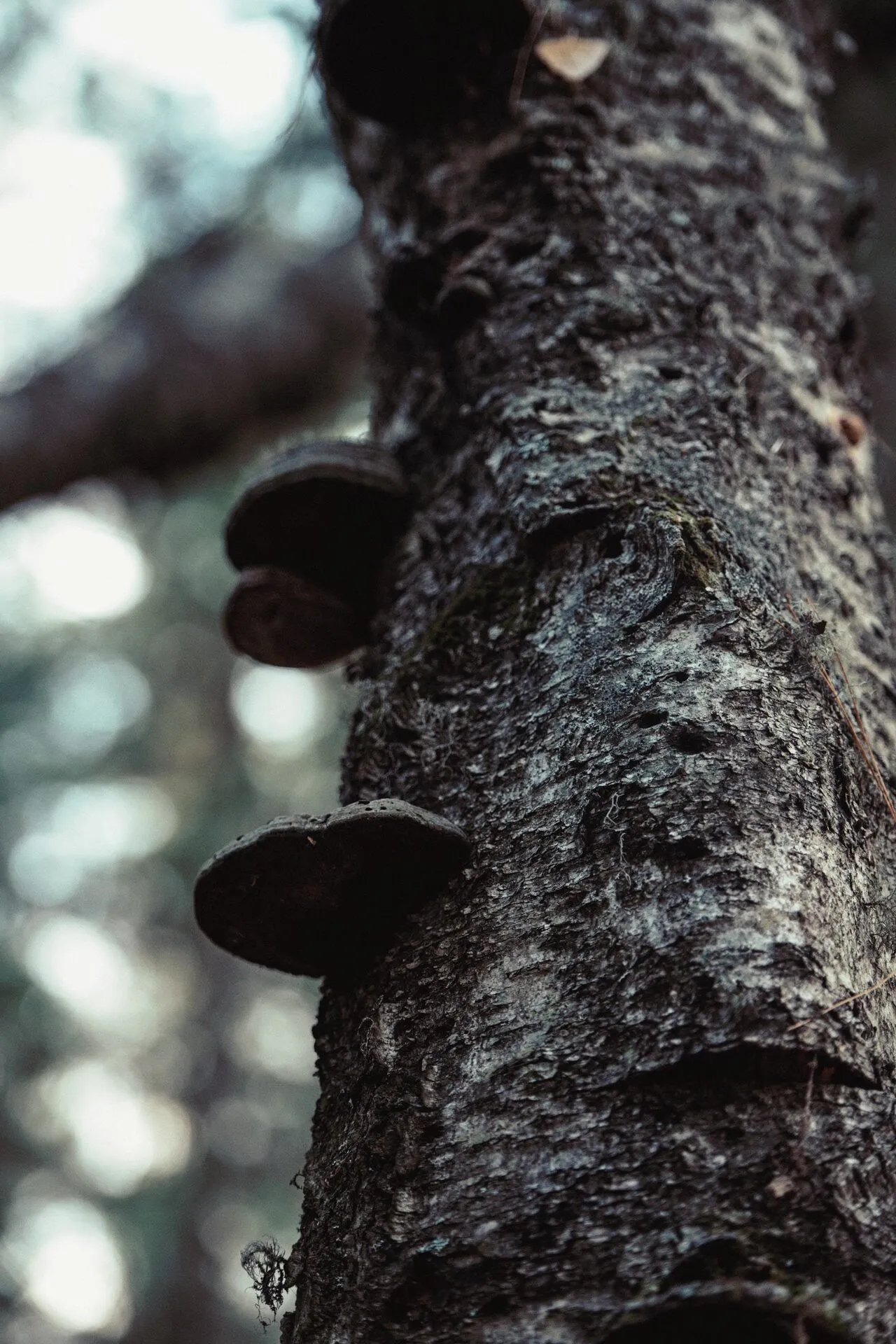
Not all mushrooms have gills. Boletes have a spongy layer of tubes and pores instead; polypores have pores; chanterelles have shallow false gills; and others have teeth, ridges, or smooth surfaces. The spore-bearing structure is one of the first things a mycologist checks.
The spacing and breadth of the gills also varies in identifiable ways. Some species carry gills crowded so densely that dozens pack into a centimeter; others are widely spaced with visible gaps. Many species have shorter gills, called lamellulae, that begin at the cap edge and stop partway toward the stem, interleaved with the full-length gills. These small consistencies — crowding, breadth, the presence of lamellulae, whether the gill edges are smooth or finely serrated — are exactly the granular features that separate species which otherwise look almost identical to a casual eye.
The color of the gills is worth watching over time. In many species the gills start pale and darken as the spores mature, eventually taking on the color of the spore print. Watching that color shift is another way to read the maturity and identity of a specimen.
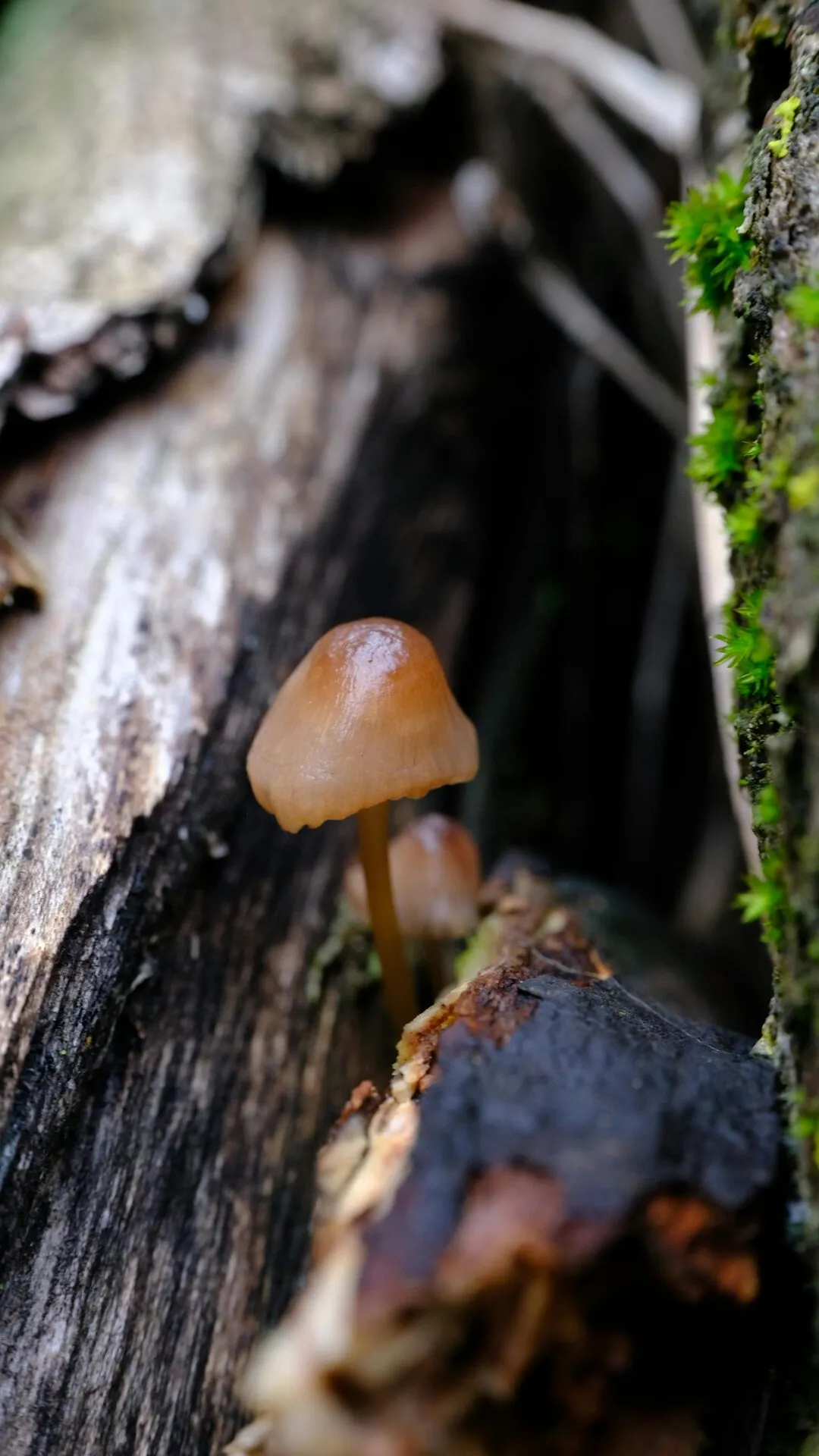
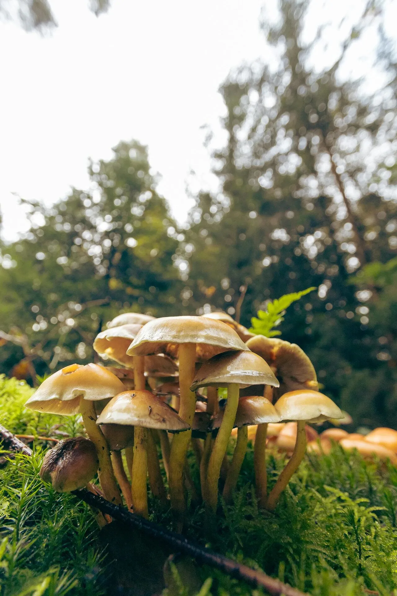
The Stem (Stipe)
The stem, or stipe, raises the cap above the substrate. This elevation is functional: getting the spore-bearing surface up into moving air dramatically improves the odds that released spores will travel away from the parent rather than falling straight back down.
Stems carry identification features of their own. They may be central, off-center, or lateral. The texture ranges from smooth to fibrous to scaly. Some are hollow, some solid, some stuffed with a cottony pith. The base of the stem is especially important — it may be equal in width, swollen into a bulb, or rooting deep into the substrate, and in some genera it carries critical structures discussed below.
In psilocybin mushrooms, the stem often shows the same blue bruising reaction as the cap, and the way it discolors when handled is part of the overall picture used in identification.
The Partial Veil and Ring (Annulus)
Many mushrooms begin life with their gills protected by a membrane called the partial veil, which stretches from the cap edge to the stem, covering the developing fertile surface. As the cap expands and matures, this veil tears.
What remains after the veil tears is often a ring of tissue around the stem, called the annulus. The ring may be a prominent skirt, a thin fragile zone, a movable band, or merely a faint fibrous line. Some mushrooms lose the ring entirely as they age.
The presence, position, and form of the ring is a significant identification feature — and a safety-critical one. Several deadly Amanita species have a distinct ring, and noticing it (along with other features) is part of distinguishing them from safer lookalikes.
The Universal Veil and Volva
Some mushrooms, most notably in the genus Amanita, begin entirely enclosed in a second membrane called the universal veil, which wraps the whole immature fruiting body like an egg. As the mushroom grows and bursts out of this envelope, remnants are left behind.
These remnants take two forms. On the cap, they may appear as patches or warts — the white flecks on a classic fly agaric are torn pieces of universal veil. At the base of the stem, the remnant often forms a cup-like structure called a volva.
The volva is one of the most important features in the entire field, because the deadliest mushrooms on Earth — the death cap and the destroying angels — have one. It is often below the soil surface, which is exactly why responsible foragers excavate the entire base of any specimen rather than cutting it at ground level. A volva missed is a clue missed, and with Amanita the stakes are lethal.
The Spore Print
The spores themselves are too small to see individually, but in mass they have color, and that color is one of the most reliable identification features available without a microscope. To reveal it, mycologists make a spore print: a cap is placed gills-down on paper or glass for several hours, and the falling spores accumulate into a visible deposit that takes on their collective color.
Spore color is far more stable than cap color. It ranges across white, cream, pink, brown, rust, purple-brown, and black depending on the species, and it often distinguishes genera that otherwise look similar. Psilocybe species, for example, characteristically produce a dark purple-brown to black spore print — a useful distinguishing feature from some toxic lookalikes that drop rusty-brown spores.
The spore print sits at the boundary between field observation and laboratory work. It requires no equipment beyond paper and patience, yet it provides information that surface appearance alone cannot.
Under a microscope, the spores themselves reveal another layer of identification features that professionals rely on. Spore shape (round, elliptical, spindle-shaped), size measured in micrometers, surface ornamentation (smooth, warted, ridged), and the presence of a germ pore — a thin spot in the spore wall where the next generation will emerge — all vary between species in consistent ways. For closely related species that cannot be separated in the field, microscopic spore features are often the deciding evidence. This is why serious identification frequently moves from the field to the bench, where a drop of mounting fluid and a microscope settle questions that the naked eye cannot.
The Hidden Body: Hyphae and Mycelium
Everything described so far is the fruiting body — the visible minority of the organism. The fungus itself is built from microscopic filaments called hyphae, which branch and fuse into the vast network known as mycelium.
A single hypha is a thread, often only a few micrometers wide, that grows at its tip and absorbs nutrients across its surface. Collectively, hyphae form an absorptive surface of staggering extent — a handful of healthy forest soil can contain kilometers of fungal filament. This is how the fungus feeds: by permeating its substrate and digesting it externally, secreting enzymes and absorbing the products.
The mushroom we see is built from the same hyphae, packed and organized into tissue. When the fungus fruits, it weaves loose filaments into the dense, structured form of cap, gills, and stem. In a sense, the mushroom is mycelium that has temporarily organized itself into a tower for the purpose of reproduction.
The hyphae are also where the fundamental biology of fungi reveals itself as distinct from both plants and animals. Fungal hyphae are typically divided into compartments by internal walls called septa, though these are often perforated, allowing cytoplasm and even nuclei to flow between compartments. The cell walls themselves are built largely of chitin — the same tough material found in insect exoskeletons — rather than the cellulose of plants. These are not surface details; they place fungi in their own kingdom, more closely related to animals than to the plants they superficially resemble. When you look at the dense white tissue inside a fresh mushroom stem, you are looking at chitin-walled hyphae packed together, the same building blocks that thread invisibly through the soil below.
Why the Structure Is Consistent
The remarkable thing about mushroom anatomy is how reliably these features recur across species. The same set of parts — cap, gills or pores, stem, sometimes a ring, sometimes a volva — appears again and again, with variations that are themselves consistent within each species.
This consistency is what makes identification possible at all. Because a given species reliably produces gills of a particular attachment, a stem of a particular texture, a spore print of a particular color, and so on, these features can be cataloged and used as a key. The variation between species is structured rather than random.
It also reflects shared evolutionary descent and shared functional pressures. The spore-delivery problem has a limited number of good solutions, and fungi have converged on the cap-and-gill architecture repeatedly because it works: it protects the fertile surface, elevates it, and maximizes spore-producing area.
Reading a Mushroom in Practice
Putting the anatomy together, an experienced observer reads a mushroom as a set of converging clues. They note the cap shape and surface, turn it over to check the spore-bearing surface and gill attachment, examine the stem and especially its base for a volva, look for a ring, take a spore print, and consider the habitat and substrate.
No single feature is sufficient. Identification — particularly identification safe enough to eat by — depends on the whole picture, with special attention to the features that distinguish dangerous species from their lookalikes. This is why casual identification from a cap photograph is unreliable and, in the case of foraging, dangerous.
For the purposes of understanding rather than foraging, the anatomy is its own reward. Once you can name the parts, a mushroom stops being a vague shape and becomes a legible structure — a record of how a hidden organism solved the problem of making more of itself.
A Vocabulary Worth Having
The terms in this guide — pileus, lamellae, stipe, annulus, volva, hymenium, hyphae, mycelium — are not jargon for its own sake. They are the working vocabulary that lets people communicate precisely about organisms that vary in subtle and consequential ways.
Most of these terms connect to the broader concepts covered across our mycology and glossary sections. The hyphae that build the fruiting body are the same filaments that form the mycelial network; the spores produced on the gills are the starting point of the fungal life cycle. Anatomy is the entry point to all of it.
Learn the parts, and the rest of mycology becomes far easier to follow. Every later topic — life cycle, ecology, classification, the distinctions between genera — builds on the structural foundation laid out here.
There is also a deeper payoff to learning this vocabulary. A named structure is a structure you can think about. Before you have the word “volva,” a cup at the base of a stem is just an odd detail; once you know what it is and which genera carry it, that same detail becomes a piece of evidence that can, in the most serious cases, distinguish a meal from a fatal mistake. Naming converts observation into knowledge. The patient work of learning anatomy is what turns looking into seeing — and in mycology, seeing clearly is the whole discipline.